Awake Craniotomy
Brain surgery where you stay awake for part of the procedure so the surgical team can map and protect speech and movement while removing a tumor.
What Is an Awake Craniotomy?
An awake craniotomy is brain surgery in which the patient is awake for part of the procedure. It is also called awake brain surgery or awake brain mapping. It is used when a tumor sits in or near areas of the brain that control speech, language, or movement. By keeping the patient awake for the part of surgery that matters most, the surgical team can test these functions in real time while the tumor is being removed.
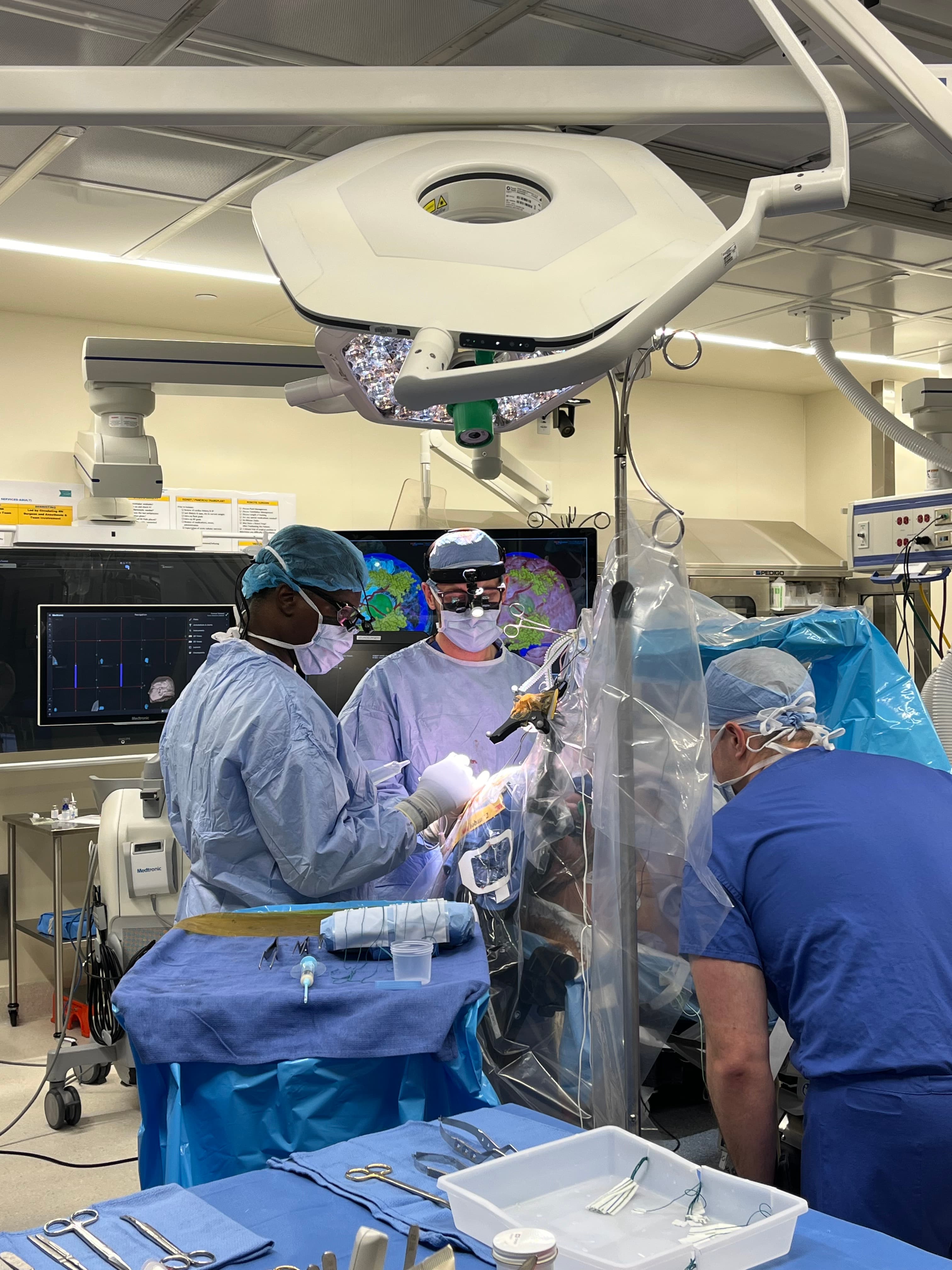
The brain does not come with a map that looks the same in every person. The exact spot that controls a word, a hand, or a sentence can sit just millimeters from a tumor, and that location varies from one person to the next. During an awake craniotomy, the surgeon applies a small electrical signal to the surface of the brain while the patient talks, counts, or moves. If a spot is tied to an essential function, the team sees the effect immediately and avoids it. This real-time feedback can allow more complete tumor removal while helping protect speech, motor function, and other abilities.
A common fear is that being awake means feeling the surgery. The brain itself has no pain receptors, so the brain tissue cannot feel pain when it is touched or operated on. The scalp, skull, and the coverings of the brain do have nerves, so those areas are numbed with a scalp block, local anesthetic placed around the nerves of the scalp. Patients are also sedated for the parts of the procedure that do not require them to be awake.
Awake craniotomy is performed by a team that includes the neurosurgeon, a neuroanesthesiologist, and specialists who guide the patient through the mapping tasks. Dr. Sherman performs awake craniotomy at Rutgers Cancer Institute in New Brunswick, NJ, the only NCI-designated comprehensive cancer center in New Jersey, and has published research on using virtual reality during awake brain mapping to test functions such as language and visuospatial cognition.
Call our office at 732-235-6333 or send us a message.
The information on this page is general educational information and is not medical advice. It does not create a doctor-patient relationship or reflect a treatment recommendation for any individual. Treatment decisions require an individual evaluation by a qualified physician.
What to Expect
The experience is more structured and calmer than most people expect. Here is the general sequence.
Consultation and mapping plan
Before surgery, the team reviews imaging and plans which functions need to be mapped based on where the tumor sits. The patient learns what tasks they will be asked to do during the awake phase, such as naming pictures or moving a hand, so nothing comes as a surprise.
Pre-op preparation
On the day of surgery, the patient is prepared in the operating room. Monitors are placed, and the neuroanesthesiologist and surgical team get everything ready. The scalp is numbed with local anesthetic around its nerves so the head can be positioned and the procedure can be done without pain.
Going to sleep first
In the common asleep-awake-asleep approach, the patient is sedated or asleep for the first part of the procedure, including the opening. They do not experience this part awake.
The awake phase and tasks
When it is time to map, the anesthesiologist lightens the sedation and wakes the patient gently. During this phase, the patient performs tasks such as counting, reading, naming objects in pictures, moving a limb, or answering questions while the surgeon stimulates and removes tumor. The patient may notice sensations like tingling or brief trouble finding a word when a spot is tested. These are expected and tell the team what to protect.
Back asleep for closure
Once the mapping and the key portion of tumor removal are complete, sedation is deepened again. The patient is asleep or sedated for the closing part of the procedure.
Recovery room
After surgery, the patient is monitored closely in a recovery area and then a hospital room. The team checks speech, movement, and overall neurologic function.
Hospital stay and home
The hospital stay is often around two to four days, depending on the tumor and how recovery goes. A first follow-up visit is commonly scheduled within a couple of weeks of surgery.
Conditions Treated with Awake Craniotomy
Awake craniotomy is used when these tumors sit in or near the brain's speech, language, or motor areas. Each condition page covers symptoms, diagnosis, and the full range of treatment options.
- Low-Grade GliomaSlow-growing gliomas that often arise in or near eloquent brain regions.
- High-Grade GliomaAggressive gliomas where mapping allows safer, more complete removal.
- GlioblastomaThe most common malignant brain tumor; awake mapping protects function during resection.
- MeningiomaSelected meningiomas near speech or motor areas can warrant mapping during removal.
- Brain MetastasesMetastases near eloquent cortex are sometimes removed with awake mapping.
Using XR Technology To Advance Medical Care
Dr. Sherman integrates VR and AR visualization into surgical planning and patient education, giving patients a clearer picture of their diagnosis and the approach to treatment.
Common Questions About Awake Craniotomy
No, the surgery itself is not felt as pain. The brain has no pain receptors, so it cannot feel pain when it is touched or operated on. The scalp and skull do have nerves, so they are numbed with local anesthetic before and during the procedure. Patients are also sedated for the parts that do not require them to be awake, and the team monitors comfort throughout.
Newly Diagnosed or Seeking a Second Opinion?
Dr. Sherman is accepting new patients at Rutgers Cancer Institute in New Brunswick, New Jersey. Whether you have just been diagnosed or you are seeking another perspective on a surgical plan, a consultation is the first step toward understanding your options.